
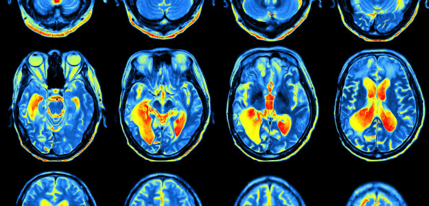
Until the recent past, brain imaging was not available to help us understand how ADHD works at the brain level. Therefore, ADHD was understood mainly by looking at behavior. Difficulty staying focused, acting impulsively, and constant movement were often the signs that led someone to seek an evaluation. But over the last two decades, advances in brain imaging have given researchers a much clearer picture of what’s happening beneath those behaviors. And what they’ve found is far more complex and more interesting than the idea of a single “ADHD brain.”
What Does ADHD Look Like in the Brain?
Brain imaging research shows that ADHD is associated with differences in brain development, connectivity, and communication across several regions involved in attention, self-regulation, motivation, and executive functioning.
Researchers have identified several brain regions that tend to develop or function differently in people with ADHD. These include:
- The prefrontal cortex – involved in planning, decision-making, impulse control, and self-regulation
- The basal ganglia – helps regulate motivation, reward, and movement
- The cerebellum – best known for coordinating movement, but also plays a role in attention and timing
- The anterior cingulate cortex – involved in monitoring mistakes, shifting attention, and selecting appropriate responses
Large brain imaging studies have found that, on average, children with ADHD tend to have slightly smaller gray matter volumes in some of these regions. In addition, there is evidence that the cortex, the brain’s outer layer which supports higher-level thinking and decision-making, may mature more slowly.
That doesn’t mean these areas are damaged or permanently underdeveloped. Instead, it suggests that, for some children, parts of the brain may simply follow a different developmental timeline. Many of these differences may become less pronounced as the brain matures.
It’s Not Just About Brain Structure
Researchers are also learning that how different parts of the brain communicate may be just as important as the size or shape of individual brain regions.
The brain’s white matter acts like a network of communication highways, carrying information between different areas. Studies have found differences in some of these connections in people with ADHD, particularly those linking the frontal lobes with deeper brain structures involved in attention, motivation, and movement.
Functional MRI (fMRI) studies add another piece to the puzzle. Unlike a standard MRI, which shows brain anatomy, an fMRI measures changes in brain activity while the brain is at work or at rest.
One network being closely studied is the default mode network. This network is most active when we’re daydreaming, thinking about ourselves, or letting our minds wander. Normally, it becomes less active when we need to concentrate on a task.
For some people with ADHD, however, this transition doesn’t appear to happen as efficiently. If the default mode network stays more active than it should during a task, staying focused can become much more difficult. While this isn’t true for everyone with ADHD, it may help explain why sustained attention often feels so mentally exhausting.
ADHD Isn’t Just One Brain Pattern
One of the biggest changes in ADHD research is the growing recognition that there may not be a single neurological profile that explains every person with ADHD.
A 2026 study published in JAMA Psychiatry analyzed brain scans from more than 1,100 children and identified three biologically distinct patterns of brain network differences, each associated with a different symptom profile:
- One pattern was linked primarily to emotional regulation.
- Another involved brain circuits responsible for action control.
- A third showed differences in networks related to sustained attention.
What’s especially interesting is that researchers found these patterns using brain imaging alone. They didn’t use symptom ratings to create the groups, yet the brain-based patterns still matched meaningful differences in behavior.
A separate 2026 study from China reached a similar conclusion using a different approach. The researchers identified two distinct brain-behavior patterns and suggested that combining everyone with ADHD into one large group may actually hide important biological differences. In short, if different subgroups show opposite brain patterns, averaging them together can make those differences disappear in research data. That may help explain why earlier imaging studies sometimes produced inconsistent findings.
Rather than weakening the evidence for ADHD, these studies strengthen it. They suggest that ADHD is a real neurodevelopmental condition with multiple biological pathways that can lead to similar symptoms…much like there are many different causes of headaches even though the symptom feels similar.
Rethinking How Stimulant Medications Work
Brain imaging research is also changing how scientists think about ADHD medications.
For many years, stimulant medications such as methylphenidate (Ritalin, Concerta) and amphetamine-based medications (Adderall, Vyvanse) were believed to work mainly by improving activity within the brain’s attention and executive function networks.
A recent imaging study from Washington University School of Medicine suggests the picture may be more complex. Using resting-state functional MRI, researchers found that stimulant medications appeared to produce some of their strongest effects in brain systems involved in arousal, motivation, and reward processing rather than exclusively in attention networks.
In practical terms, this means the medications may help people focus not simply by “turning on” attention, but by making it easier for the brain to stay engaged with tasks that might otherwise feel tedious or unrewarding.
That doesn’t mean scientists have completely figured out how stimulants work, or that attention networks aren’t involved. ADHD affects multiple interconnected brain systems, and stimulant medications likely influence several of them at the same time. Researchers are continuing to study how these brain changes relate to improvements in everyday functioning.
What This Research Does, and Doesn’t, Tell Us
As exciting as these discoveries are, it’s important not to overinterpret them.
Brain scans cannot diagnose ADHD. Diagnosis is still based on a careful clinical evaluation that includes symptom history, interviews, and standardized rating scales.
The brain differences described here are findings seen across large groups of people. They don’t yet provide a reliable way to determine whether one individual has ADHD or to identify which biological pattern they may fit into.
What decades of research do show is that ADHD has a measurable neurobiological basis. Across many large studies using different imaging techniques, researchers have consistently found differences in brain development, connectivity, and function.
The next challenge is figuring out how to use this knowledge. If future research can match specific brain patterns with the treatments most likely to help, ADHD care may eventually become much more personalized than today’s trial-and-error approach.
The Bottom Line
Brain imaging has transformed our understanding of ADHD. Rather than pointing to a single brain circuit, research suggests that ADHD involves several different patterns of brain development and communication that can lead to similar symptoms.
There’s still much to learn, but each new study brings researchers closer to understanding why ADHD looks different from one person to another and how treatment may someday become more individualized as a result.
What Does This Mean for Treatment?
Although brain imaging has expanded our understanding of ADHD, treatment decisions are still based on a person’s symptoms, strengths, daily challenges, and goals – not on brain scans.
Evidence-based treatment may include behavioral therapy, school or workplace accommodations, medication when appropriate, and healthy lifestyle habits such as regular exercise and good sleep. Some people also choose to incorporate complementary approaches, including neurofeedback, as part of a broader treatment plan.
As research continues to uncover the different brain networks involved in ADHD, the hope is that future treatments will become increasingly personalized, helping clinicians match individuals with the approaches most likely to benefit them.
Ready to Learn More About ADHD Treatment?
Understanding how ADHD affects the brain is an important first step, but effective treatment is always about the individual and not just the diagnosis. Every person with ADHD has unique strengths, challenges, and goals, which is why a personalized approach is so important.
At Northwest Neurofeedback, we provide comprehensive neurofeedback assessments and individualized training programs designed to support attention, focus, self-regulation, and executive functioning. Whether you’re exploring options for yourself or your child, we’re here to help you understand the evidence, answer your questions, and determine whether neurofeedback may be an appropriate part of your treatment plan.
If you’d like to learn more, contact us to schedule a complimentary 15-minute consultation and discover how a personalized approach may help you or your child thrive, or visit https://chadd.org and https://add.org
This article summarizes findings from peer-reviewed research and is intended for educational purposes only. It is not a substitute for professional medical advice, diagnosis, or treatment. If you have questions about ADHD or believe you or your child may have ADHD, consult a qualified healthcare provider.